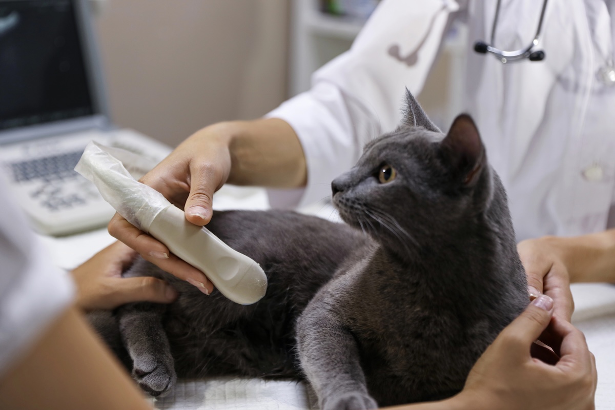
The Basics On Ultrasound for Pets
Ultrasound for pets – aka Ultrasonography – has had a major impact on how we practice veterinary medicine. It provides a non-invasive means of looking inside our patients, greatly improving how we approach medical cases from a testing and treatment standpoint.
Ultrasound For Pets: How Does Ultrasonography Work?
An ultrasound machine produces high frequency (ultrasonic) sound waves that are pulsed through a probe placed against a patient’s skin. Ultrasonic sound waves echo back to the probe as they encounter different structures within the body. Sound interacts with tissues and substances differently depending on their individual characteristics. This, in turn, creates differences in the echoes returning to the probe. A computer uses these differences to create the image that appears on the ultrasound machine’s screen.
Many ultrasound machines also have Doppler functionality. Doppler makes use of sound interacting with blood flowing through the heart and blood vessels to give us information regarding the speed and direction of the blood flow.
Ultrasound for Pets: What are the Advantages of Ultrasound?
To understand the advantages of ultrasonography, we need to compare it to other techniques for evaluating the inside of our patients.
Diagnostic radiography makes use of X-rays passing through the body and striking a plate or film to create an image called a radiograph. These X-rays are a form of radiation that has the potential to harm the body’s tissues. Our patients are at minimal risk: most pets have no more than a handful of radiographs taken during their lives, and we use a technique chart to ensure that only the necessary level of radiation is used over only the specific body part we’re evaluating. There is a greater risk to the staff members who are repeatedly radiographing numerous pets each week. Shielding, appropriate technique, and monitoring of staff exposure levels help minimize these risks, but they’re not zero.
Ultrasound imaging eliminates this risk—there is NO harmful radiation for people or pets.
Radiographs also are two-dimensional, still images. Ultrasound allows us to evaluate internal structures in real time and to quickly rotate the probe to see the body from different angles.
Advanced imaging techniques like computed tomography (CT) and magnetic resonance imaging (MRI) require referral to a specialized facility and general anesthesia. At Alford Avenue Veterinary Hospital, ultrasound procedures can be performed in-house and typically require no anesthesia.
Ultrasound for Pets: Ideal Applications of Ultrasound
Assessment of Organs: Ultrasound can be used to briefly evaluate a single body structure or organ like the urinary bladder. We frequently use ultrasound to determine if a patient’s urinary symptoms might be caused by a bladder stone or to determine if the bladder is full enough to allow for collection of a urine sample for testing.
Pregnancy Determination: Ultrasound can be used to determine whether a patient is pregnant and to assess the general health of the fetuses. In dogs and cats, pregnancy can be determined as early as 25 days after fertilization.
Internal Medicine Diagnoses: In the case of a sick or injured pet, point-of-care ultrasound can be used to quickly and accurately look for internal bleeding; fluid accumulation in the abdomen, lungs, or chest cavity; signs of anaphylactic shock; or obvious masses.
Abdominal Assessments: A full abdominal ultrasound is used to systemically scan the entire abdominal cavity, evaluating the appearance of the organs and making measurements to compare to references. Such a scan is often indicated for patients with lab work abnormalities that point to organ dysfunction, patients with chronic gastrointestinal symptoms, or patients with poor appetite and/or weight loss, among others.
Assessment of Cardiac Issues: Echocardiography is used to evaluate the size, structure, and function of the heart and its valves. These scans allow us to determine if clinically significant heart disease is present, whether any medication is needed, or whether previously prescribed medication is working as intended.
Ultrasound For Pets: How To Schedule An Ultrasound Procedure
If we determine that your pet needs an ultrasound, we often can perform imaging on the same day as his or her visit. Echocardiograms are most often scheduled in advance, and we will find a time that works for you before you leave your appointment if a same day scan isn’t feasible.
If Alford Avenue Veterinary Hospital is not your pet’s primary clinic, we also accept referrals and will coordinate with the doctor(s) at your home clinic regarding results and treatment recommendations. We understand that specialty clinics and universities can have long wait times for appointments. If we can offer some answers sooner, we’re always happy to help, though there are times that referral to a specialty center remains the best option.
Seeking an ultrasound for pets in the Birmingham-Hoover, Alabama area? Alford Avenue Veterinary Hospital provides expert ultrasound diagnostics. In 2025, the hospital installed a new, state-of-the-art ultrasound machine that offers higher-resolution images.
Contact Us for a consult or to schedule an ultrasound for your pet.